Extensor Pollicis Brevis (EPB) Anatomy
- Origin: Radius (dorsal surface of the distal third of the radius inferior to the origin of the EPL) and the adjacent interosseous membrane
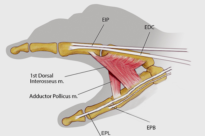
- Insertion: Thumb MP joint dorsal joint capsule and the dorsal base of the proximal phalanx of the thumb
- Innervation: Cervical roots - C7 and C8
- Nerve: Radial nerve (posterior interosseous branch)
Diagrams & Photos
Videos
Extensor Pollicis Brevis (EPB)
Key Points
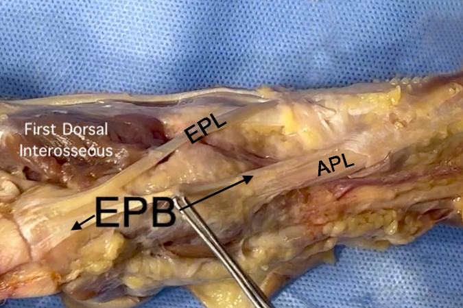
- The extensor pollicis brevis is a a tiny muscle/tendon unit which originates in the dorsal forearm and passes through the first dorsal extensor compartment to reach its insertion site on the dorsal thumb.
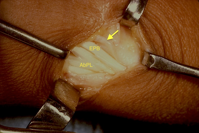
- Inside the first extensor compartment the EPB's single slip usually has a separate unique compartment dorsal and ulnar to the EPL. When treating DeQuervain's tenosynovitis surgically, this interior separate compartment must be identified and released separately during any first extensor compartment tendon sheath release surgery.


