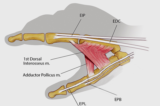
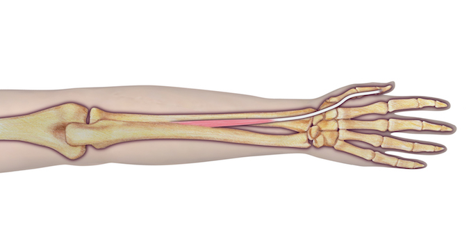
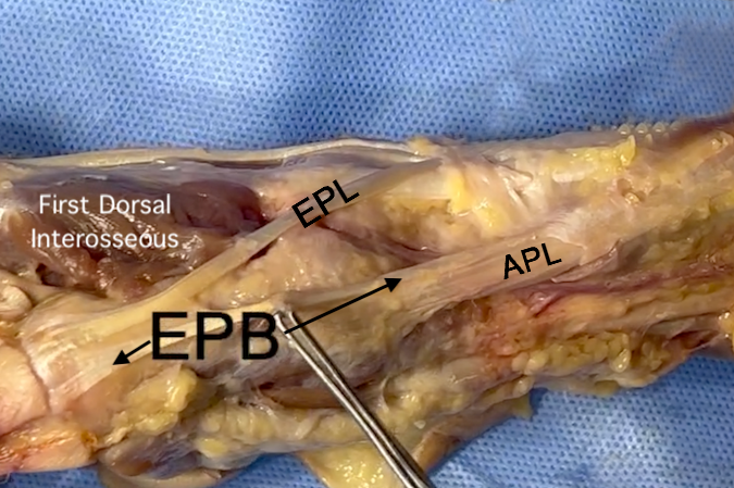
Extensor Pollicis Longus (EPL) Anatomy
- Origin: Ulna (posterolateral surface of middle shaft) Interosseous membrane
- Insertion: Thumb (base of distal phalanx, dorsal side)
- Innervation: Cervical roots: C7 and C8
- Nerve: Radial nerve (posterior interosseous branch)
Diagrams & Photos
Videos
EPL, EPB & APL Video
Key Points
- The extensor pollicis longus (EPL) usually retracts vigorously after laceration proximal to its connections to the extensor hood at the thumb MP joint.
- The retracted extensor pollicis longus (EPL) can develop a contracture of the muscle tendon complex very quickly, therefore, an early repair is essential.
- Late treatment for a neglected extensor pollicis longus (EPL) laceration will frequently require reconstruction with a EIP tendon transfer.


